Brain Tumor Segmentation with Deep Learning.
Physiology-informed brain tumor segmentation using DSC MRI hemodynamic properties and learning-based image analysis.
Project
- — Deep Learning
- — Medical Imaging
- — Brain Tumors
Notes
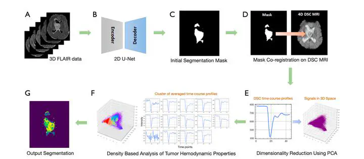
This project explores how DSC MRI hemodynamic information can add physiological context to brain tumor segmentation. By combining structural segmentation with density-based analysis of perfusion time courses, the method highlights tissue behavior that conventional morphology-only segmentation can miss.
Project Overview
Brain tumor segmentation is a critical task in medical imaging, especially when tumor tissue is heterogeneous and difficult to characterize from structural scans alone. This project addresses that problem by:
- Integrating hemodynamic data with traditional imaging modalities
- Combining U-Net segmentation with density-based analysis of DSC MRI time courses
- Adding physiological context to segmented regions for more interpretable image analysis
Key Contributions
- Incorporation of DSC MRI hemodynamic properties into a segmentation workflow
- More physiologically informed characterization of heterogeneous tumor regions
- A framework for comparing structural segmentation with perfusion-derived tissue behavior
Research Impact
This work contributes to AI-assisted medical imaging and has potential applications in:
- Clinical research: Physiological context for tumor boundary interpretation
- Treatment planning: Better visualization for surgical and radiation therapy
- Research: Enhanced understanding of tumor hemodynamics
The research has been published in Quantitative Imaging in Medicine and Surgery and represents a step toward segmentation workflows that account for both image structure and tissue physiology.