Brain Tumor Segmentation with Deep Learning
 Brain tumor segmentation visualization
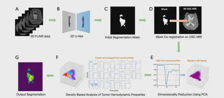
This project focuses on developing advanced deep learning techniques for automated brain tumor segmentation in medical imaging. By incorporating hemodynamic properties into the segmentation process, we achieve improved accuracy in identifying tumor boundaries and characteristics.
Project Overview
Brain tumor segmentation is a critical task in medical imaging that directly impacts treatment planning and patient outcomes. Traditional segmentation methods often struggle with the complex and heterogeneous nature of brain tumors. This project addresses these challenges by:
- Integrating hemodynamic data with traditional imaging modalities
- Developing novel deep learning architectures optimized for medical image analysis
- Improving segmentation accuracy for better clinical decision-making
Key Contributions
- Novel incorporation of hemodynamic properties in deep learning models
- Enhanced segmentation performance on challenging brain tumor cases
- Potential for clinical translation and improved patient care
Research Impact
This work contributes to the growing field of AI-assisted medical imaging and has potential applications in:
- Clinical diagnosis: More accurate tumor boundary detection
- Treatment planning: Better visualization for surgical and radiation therapy
- Research: Enhanced understanding of tumor hemodynamics
The research has been published in Quantitative Imaging in Medicine & Surgery and represents a significant step forward in applying artificial intelligence to medical imaging challenges.
Rory
PhD Student
I am a PhD student in Biomedical Engineering at Duke University, supervised by Dr. Dan Ma. I recently completed dual majors in Applied Mathematics and Biology at Emory University. My research interests focus on quantitative MRI and deep learning for medical imaging, especially MR fingerprinting and reconstruction. Feel free to reach out at any time!